Breast Imaging

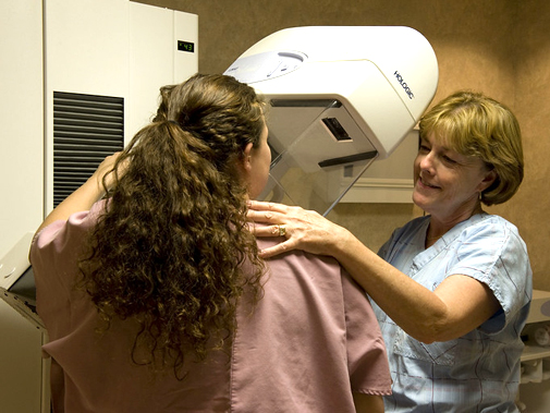
Annual screening mammogram
All mammography machines at our facilities use digital technology. With digital mammography, the radiologist reviews electronic images of the breast using special high-resolution monitors. The physician can adjust the brightness, change contrast and zoom in for close ups of specific areas of interest.
We follow the guidelines of the American Cancer Society, which recommends women begin yearly mammograms at age 40. In addition to yearly mammograms, women 40 and older should also get a breast exam by a healthcare professional every year (women in their 20s and 30s should have a breast exam at least every three years). Patients with a first-level family history of breast cancer that developed before menopause should talk with their physicians, who may recommend starting mammograms at a younger age and seeking additional risk assessment and consultation with our breast experts.
Breast MRI services
A breast MRI uses magnetic fields to create an image of the breast. MRI can help distinguish between benign (noncancerous) and malignant (cancerous) areas. During the procedure, a dye is injected through a vein in the arm. The dye makes a tumor much brighter than the surrounding breast tissue and visible on the MRI scan. Although MRI can detect tumors in dense breast tissue, it cannot detect tiny specks of calcium (known as microcalcifications), which account for half of the cancers detected by mammography. MRI can be a good tool for breast cancer screening when used together with mammography in certain groups of women at higher risk.
Tomosynthesis (3D mammography)
Tomosynthesis is a new type of mammogram technology used at Salem Hospital. Also called 3D mammography, it takes images from multiple angles and then builds a 3D image that a radiologist can manipulate, providing a clearer, more accurate view of the breast. Learn more
Diagnostic mammogram
A diagnostic mammogram is problem-solving exam for patients who have abnormal breast symptoms, a history of breast cancer, who have breast implants, or who have had a screening mammogram that has shown suspicious or abnormal areas needing further review. Diagnostic exams usually take 30 minutes and provide additional breast views for a more comprehensive examination. A radiologist specializing in breast imaging is on site and reviews and interprets exams while patients wait. If a suspicious area is detected, patients can be scheduled for a follow-up appointment or diagnostic procedure within 48 hours.
Diagnostic breast ultrasound
Using high-frequency sound waves, a physician can evaluate palpable breast lumps to determine whether they are fluid-filled cysts (not cancer) or solid masses (which may or may not be cancer). This technology may be used along with mammography.
High-risk management program
For women at risk for breast cancer (including those with a first-level relative diagnosed with breast cancer before menopause), we offer a high-risk management program. Women with breast cancer symptoms, or whose screening mammogram shows suspicious results, can be referred by a physician for further diagnostic testing. If necessary, patients may also be referred to us for a breast surgery consultation and comprehensive cancer treatment. Our experts are also a resource for second opinions for patients considering treatment options.

